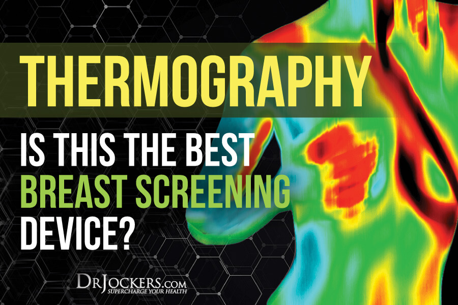
Thermography: Is This the Best Breast Screening Device?
Thermography is a high-technology tool that specifically measures inflammation in the body. This test is particularly good for assessing areas of inflammation and increased metabolic rate. It is more effective and is significantly less invasive than mammography.
Research has shown that the major mechanism involved in all degenerative diseases is inflammation. Most medical testing searches for disease processes that have already developed. They are looking downstream to the effect rather than upstream at the underlying cause. More advanced health care practitioners use instruments and technology that search upstream for the cause of physiological abnormalities in the body.

How Does Thermography Work?
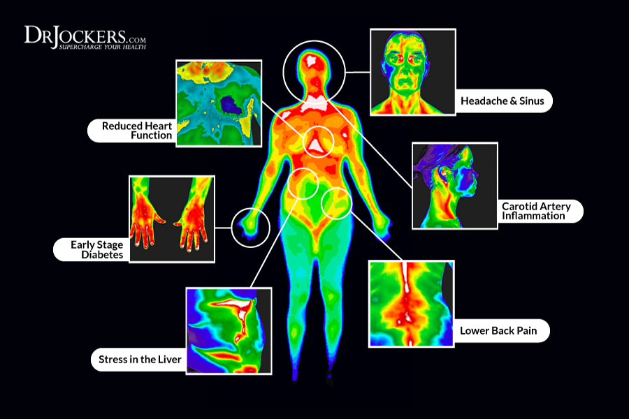
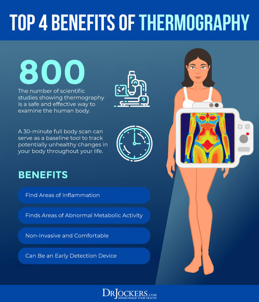
Thermography is a scanning device that measures your body surface temperature and presents the information as a digitized image. This tool makes a digital map of your body and very accurately illustrates heat patterns. These patterns may detect some abnormal condition, such as cancer cell growth or active infection.
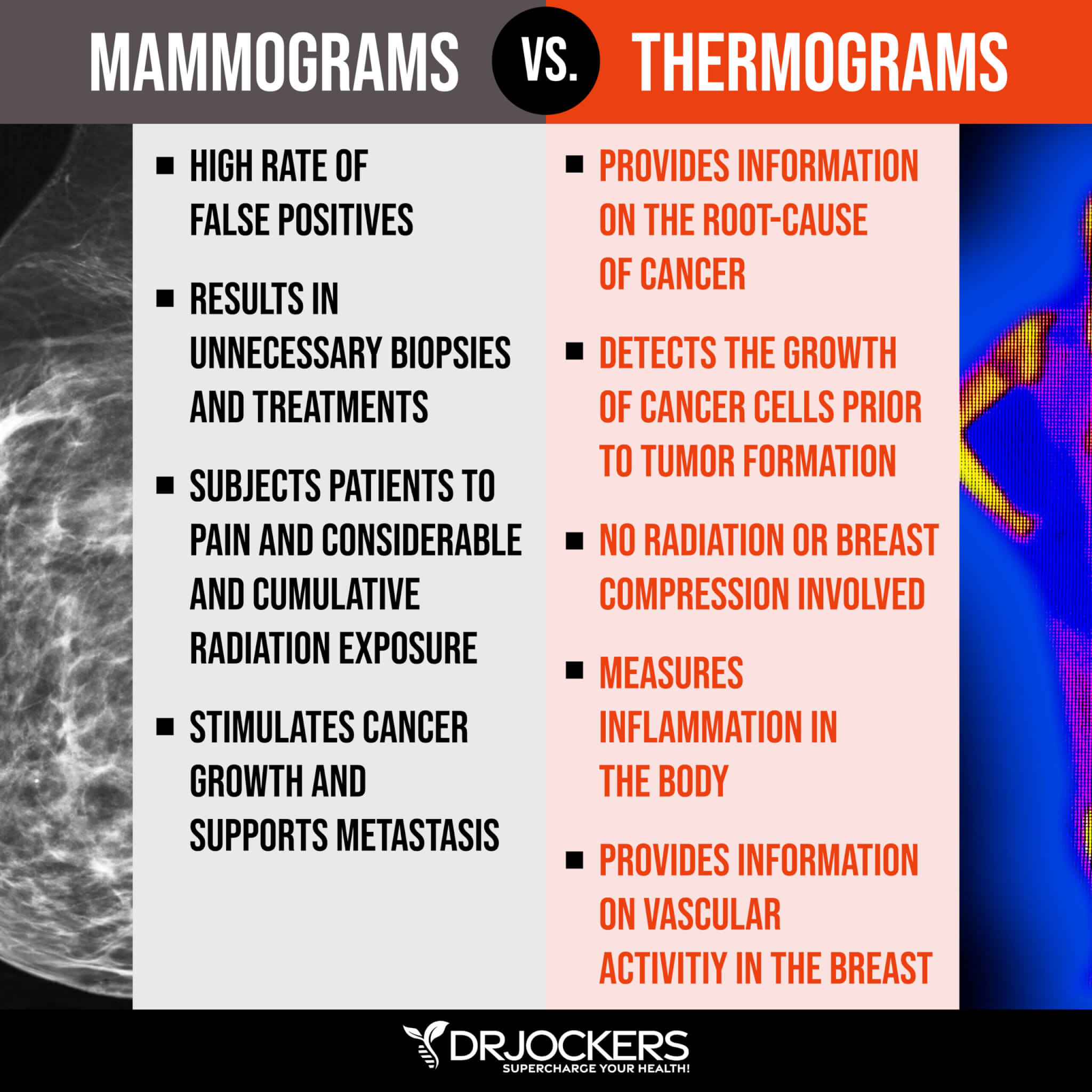
Mammograms look for anatomical changes in the breast, such as masses or lumps. Thermograms analyze the vascular changes in the breast. Increased blood into certain regions of the body increases the heat of that region. Areas of inflammation, cancer cell formation, and active infection have elevated circulation. Thermal imaging has a great ability to detect subtle physiological changes that accompany pathology (1).
Thermal Asymmetry Indicates Problems:
The body should naturally have thermal symmetry. Areas of asymmetry can indicate problems and are analyzed specifically for underlying pathology. Cancer cells divide very rapidly and demand increased blood flow and nutrient delivery.
The metabolic processes in the body cannot differentiate between cancer cells and healthy cells. This results in increased formation of blood cells around these active cancer cells.
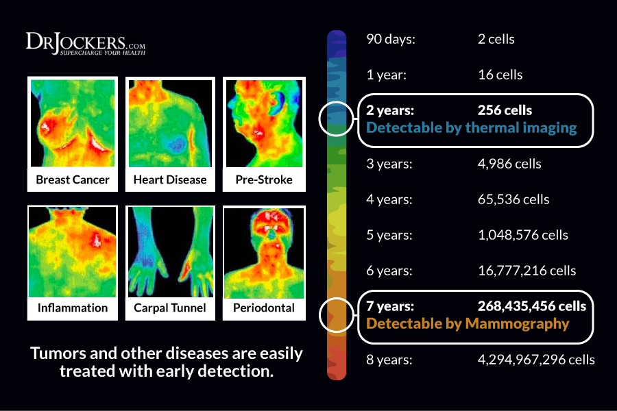
Thermography picks up this abnormal blood supply well before the cancer gets large enough to be noticed as a lump in a breast exam. It is estimated that thermography can detect cancer formation roughly 10 years before mammography can identify a tumor (2).
Breasts Normally Appear Purple:
The breasts do not normally generate much heat. In fact, healthy breasts appear purple during a thermographic exam. This indicates very low heat levels. Red, orange, or yellow spots that appear during a breast thermograph may indicate the presence of cancer and should be looked at more closely (3).
Thermography has been studied in detail for over 30 years. The database includes over 250,000 women who have been included as study participants (4). These large, long-durational studies have shown an average sensitivity and specificity of 90%. The studies show that a persistent abnormal thermogram carries with it a 22-time higher risk of future breast cancer (5).
Thermograms are Very Reliable:
Thermograms are a very reliable and accurate tool that provides precise and objective data on thermal information. This information can be used for successful diagnosis, treatment, & prognosis. They are completely painless, non-invasive, and take less than 15 minutes for a thorough reading.
Unlike mammograms, thermograms emit no harmful ionizing radiation. Mammograms are one of the more dangerous medical tools due to the very high amounts of ionizing radiation (6). Thermograms use infrared technology that is completely safe.
They also do not compress the breast tissue like mammograms. This compression that takes place during mammograms can cause cancer cells to break off and create a malignant spread through the bloodstream (7).
Thermography Testing Protocols:
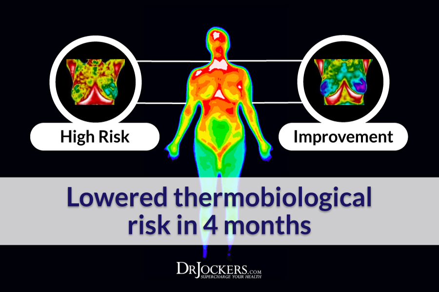
The first session one receives provides the baseline reading. Many practitioners call this the `thermal signature.` A second reading is typically recommended 3 months later to test for any changes. After these initial 2 patterns are analyzed, the patient is recommended to receive yearly thermographs to detect any subtle changes in vascularity and blood flow dynamics.
If you are outside of Atlanta, Google search thermography in your area, and you will most likely find clinics in every major city that offer this form of testing.
Inflammation Crushing Ebundle
The Inflammation Crushing Ebundle is designed to help you improve your brain, liver, immune system and discover the healing strategies, foods and recipes to burn fat, reduce inflammation and thrive in life!
As a doctor of natural medicine, I have spent the past 20 years studying the best healing strategies and worked with hundreds of coaching clients, helping them overcome chronic health conditions and optimize their overall health.
In our Inflammation Crushing Ebundle, I have put together my very best strategies to reduce inflammation and optimize your healing potential. Take a look at what you will get inside these valuable guides below!

Sources For This Article Include:
1. Institute for the Advancement of Medical Thermology Link Here
2. Sterns EE, Zee B, SenGupta S, Saunders FW. Cancer. Thermography. Its relation to pathologic characteristics, vascularity, proliferation rate, and survival of patients with invasive ductal carcinoma of the breast. 1996 Apr 1;77(7):1324-8. PMID: 8608510
3. Head JF, Wang F, Elliott RL. Breast thermography is a noninvasive prognostic procedure that predicts tumor growth rate in breast cancer patients. Ann N Y Acad Sci. 1993 Nov 30;698:153-8. PMID: 8279754
4. Breast Thermography Link Here
5. United Breast Cancer Foundation Breast Thermography Link Here
6. Heyes GJ, Mill AJ, Charles MW. Mammography-oncogenecity at low doses. J Radiol Prot. 2009 Jun;29(2A):A123-32. PMID: 19454801
7. van Netten JP, Cann SA, Hall JG. Mammography controversies: time for informed consent? J Natl Cancer Inst. 1997 Aug 6;89(15):1164-5. PMID: 9262257


Please site your peer reviewed research. People’s lives are at stake.
From the American Cancer Society:
Thermography has been around for many years, but studies have shown that it’s not an effective screening tool for finding breast cancer early. Although it has been promoted as helping detect breast cancer early, a 2012 research review found that thermography detected only a quarter of the breast cancers found by mammography. Thermography should not be used as a substitute for mammograms.
SF,
The American Cancer Society is in the cancer business. I wouldn’t trust their standards on the matter. They have the same solutions they have had for years – chemotherapy and radiation, and yet, the body heals inside out – and they’ve ignored the natural answers for years. Why? Because the cancer business is to lucrative. Follow the money.
“WE WERE SUDDENLY SEEING 20% OF THE CANCERS DIAGNOSED WERE DUCT CARCINOMA IN SITU. BEFORE [MAMMOGRAPHY] SCREENING, WE HARDLY EVER SAW THIS CONDITION. IT’S NOT A CLINICAL CONDITION, IT’S A RADIOLOGICAL CONDITION.”
–Professor Emeritus of Surgery, British Surgical Oncologist
DR. MIKE BAUM
(KING’S COLLEGE LONDON, UNIVERSITY COLLEGE LONDON)
Skip to 9:09 THE PROMISE (DOCUMENTARY ON THE HARMS OF BREAST MAMMOGRAPHY)
https://www.bitchute.com/video/iTweBm61b57M/
NOT BREAST CANCER-CAUSING MAMMOGRAPHY!
THERMOGRAPHY + ULTRASOUND OR MRI
should be the new standard for breast cancer detection and monitoring (96% sensitivity, and 98% specificity).
Integrative medicine physician Dr. Ben Johnson, M.D. at the 42nd Annual Cancer Convention by the Cancer Control Society shares a breast cancer patient’s THERMOGRAPHY results COMBINED WITH ULTRASOUND, EXTREME ALKALINE DIET, EXTREME ALKALINE FOODS, and INSULIN-POTENTIATED THERAPY (VERY LOW-DOSE CHEMO).
https://www.youtube.com/watch?v=OvPViqbnd7Y
Thanks for sharing!
When I started the Knoxville Comprehensive Breast Center 32 years ago, I did do breast thermography along with mammography and breast ultrasound. I did so for at least 3 to 5 years. I compared and correlated the results of thermography with its early breast cancer detection abilities with the other two breast imaging studies . At that time, thermography was a mylar plate that was placed on the breasts until the plate filled with colors and a Polaroid picture was taken. I can confidently say that breast thermography does not find early breast cancer. That is what we have to do- find it early if it exists in order to survive the disease. Breast thermography was initially incorporated into the BCDDP study funded by the ACS and NCI. It was conducted between 1973 and 1980. The study screened 283,000 women annually for 5 years with mammography and breast.thermography. After 2 years, breast thermography was eliminated from the study as it did not find early breast cancer.
The NCI current guidelines state that breast thermography is not for breast screening. Those facilities that incorporate this technique are not radiologists who are dedicated breast imagers. They have never correlated its results with mammography or breast ultrasound screening. I do believe that the only reason people are fascinated with it is they have made it a more sophisticated looking study with fancy electronics, but still is no different from the thermography used 35-40 years ago.
Dr. Kozlowski
Medical Director of KCBC
Clinical Breast Radiologist
The latest study confirms that mammograms do not save lives but do lead to over-diagnosis.
https://www.bmj.com/content/348/bmj.g366
I would not trust the NCI any more than I could trust the CDC to investigate the cause of autism. How can anyone compare the technology for thermography between now and forty years ago? Surely there have been advances with more sophisticated equipment? I don’t trust mammograms. I’ve only ever had one and I will never have another one. Rather work on prevention, self-checking and thermography or even sonogram. I certainly don’t trust any cancer institute or any data they publish. Most of their income goes on wages/marketing and any research they do is tied in to Big Pharma and toxic and deadly treatments. No thank you!
need to find a place to do this near Port Charlotte Fr.
Hi Susan, you can use this site to help find a clinic near you: https://www.thermologyonline.org/Breast/breast_thermography_clinics.htm
Are there any clinics in England thank you
I am sure there are. I would do a google search for them.
I try to refrain from commenting on posts such as this but my inner voice could not let this one go. First off it is very frustrating to hear anyone comparing mammography to thermography. Because the two technologies complement one another they should not be considered an alternative or replacement for one another. Mammograms are looking for a mass that has already formed. Thermography is looking for what happens prior to the mass forming. As a preventative tool we now have a way to measure current and future risk for breast disease. The technology of thermal imaging has changed drastically over the last 30 years. The radiologist in the comments above states the high risk thermograms performed in the BCDDP study from 1973 to 1979 were incorrect. What she doesn’t go on to say is that the follow-up study performed on these high risk thermograms several years later, 80% of those high risk thermograms ended up with breast cancer. The reality is that neither of these two technologies has a 100% detection rate for breast disease or cancer. Thermography is FDA approved as an adjunct to mammography, ultrasound and MRI. It should also be noted that the technology is not currently regulated so the public needs to be aware of centers that are recognized for high standards of care and FDA medically approved infrared equipment. There are four organizations currently that are recognized. The International Academy of Clinical Thermology, International Thermographic Society, American Academy of Medical Infrared Imaging and American Academy of Thermology.
Images should always be done in grayscale and color. Grayscale images provide the vascular component needed to evaluate unusual patterns ( arteries and veins) and excessive estrogen stimulation. Unfortunately many thermography centers are doing imaging in color only. http://Www.iact-org.org for more information.
Thanks for sharing Karla!
Is this available in Maryland? How much does this typically cost, I do not have insurance?
Hey Sarah, This website will help you locate one near you!